Catheters / Tubes / Implanted Devices
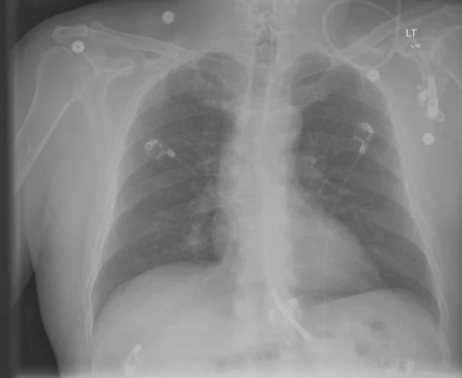
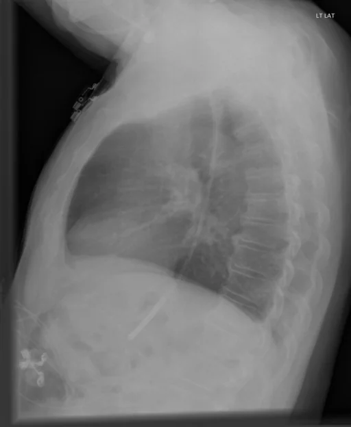
What does this chest radiograph show?
This chest radiograph is indexed in the Indiana University Chest X-ray Collection under “Catheters / Tubes / Implanted Devices”. The principal recorded finding is “Catheters / Tubes / Implanted Devices”, and the case status is documented as “Active or unspecified chronicity”. The structured record behind this page carries a single indexed term; in the source report text, a findings section, an impression and a comparison study are all present. The study comprises 2 views.
This summary is composed from the collection's structured annotation fields, and is not a description of the image.
- Diagnostic category: Catheters / Tubes / Implanted Devices
- Imaging coverage: 2 views, native resolution 512x420 pixels
- Related keywords: Catheters / Tubes / Implanted Devices, Tube, Inserted, Chest radiograph and Indiana University Chest X-ray Collection
Radiologist's Diagnostic Impression (analytical summary)
Recorded findings: Catheters / Tubes / Implanted Devices and Tube, Inserted. Categories: “Catheters / Tubes / Implanted Devices”. Status: “Active or unspecified chronicity”.
Case Radiographs (2 Views)
Radiographic Study Specifications
- Case Number
- CXR 1687
- Modality
- Chest Radiography (X-Ray)
- Views
- 2 images
- Clinical Status
- Active or unspecified chronicity
Primary Clinical Report (verbatim source text - NoDerivatives)
- Indication
- Adult male, dysphasia.
- Comparison
- PA and lateral views of the chest.
- Findings
- Feeding tube noted with tip distal to the gastroesophageal junction. No focal areas of consolidation. Heart size within normal limits. No pleural effusions. Osseous structures intact.
- Impression
- 1. No acute cardiopulmonary abnormality. 2. Feeding tube tip distal to the gastroesophageal junction.
Related Radiology Cases
Source, Scientific Provenance and Licence
Indiana University Chest X-ray Collection · Open-i / U.S. National Library of Medicine · CC BY-NC-ND 4.0
Licence: CC BY-NC-ND 4.0
Primary Data Source: https://openi.nlm.nih.gov/