Pulmonary Fibrosis / Interstitial Lung Disease and Markings (bronchovascular)
What does this chest radiograph show?
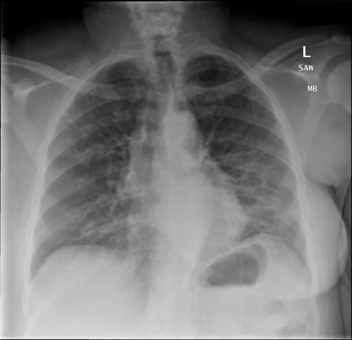
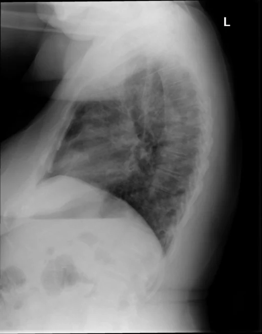
This chest radiograph is indexed in the Indiana University Chest X-ray Collection under “Pulmonary Fibrosis / Interstitial Lung Disease and Markings (bronchovascular)”. The principal recorded finding is “Pulmonary Fibrosis / Interstitial Lung Disease”, and the case status is documented as “Active or unspecified chronicity”. Recorded descriptors for this case include severity Diffuse. The structured record behind this page carries two indexed terms; in the source report text, a findings section, an impression and a comparison study are all present. The study comprises 2 views.
This summary is composed from the collection's structured annotation fields, and is not a description of the image.
- Severity and descriptors: Diffuse
- Diagnostic category: Pulmonary Fibrosis / Interstitial Lung Disease and Markings (bronchovascular)
- Imaging coverage: 2 views, native resolution 512x495 pixels
- Related keywords: Pulmonary Fibrosis / Interstitial Lung Disease, Markings (bronchovascular), Markings, Pulmonary Fibrosis, Lung and Interstitial
Radiologist's Diagnostic Impression (analytical summary)
Recorded findings: Pulmonary Fibrosis / Interstitial Lung Disease, Markings (bronchovascular), Markings (Lung, Interstitial, Diffuse) and Pulmonary Fibrosis. Categories: “Pulmonary Fibrosis / Interstitial Lung Disease and Markings (bronchovascular)”. Status: “Active or unspecified chronicity”.
Case Radiographs (2 Views)
Radiographic Study Specifications
- Case Number
- CXR 2158
- Modality
- Chest Radiography (X-Ray)
- Views
- 2 images
- Clinical Status
- Active or unspecified chronicity
Primary Clinical Report (verbatim source text - NoDerivatives)
- Indication
- The patient is an Adult female with history of pneumonia, upper lobe crackles. Known history of UIP.
- Comparison
- CT, chest
- Findings
- There are diffuse increased interstitial markings, suggestive of pulmonary fibrosis in bilateral lung fields. The fibrosis appears to slightly increased compared to previous examination, in. The trachea is midline. Negative for pneumothorax, pleural effusion. The heart size is normal.
- Impression
- 1. Redemonstrated pulmonary fibrosis without evidence for acute infiltrate.
Related Radiology Cases
Source, Scientific Provenance and Licence
Indiana University Chest X-ray Collection · Open-i / U.S. National Library of Medicine · CC BY-NC-ND 4.0
Licence: CC BY-NC-ND 4.0
Primary Data Source: https://openi.nlm.nih.gov/