Pulmonary Atelectasis and Density
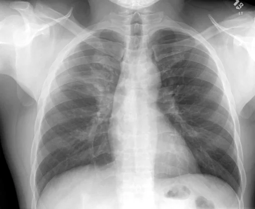
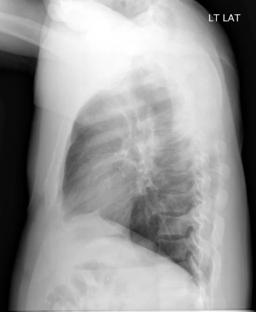
What does this chest radiograph show?
This chest radiograph is indexed in the Indiana University Chest X-ray Collection under “Pulmonary Atelectasis and Density”. The principal recorded finding is “Pulmonary Atelectasis”, and the case status is documented as “Active or unspecified chronicity”. Recorded descriptors for this case include laterality Right, and severity Mild. The structured record behind this page carries two indexed terms; in the source report text, a findings section and an impression are present, with no comparison study recorded. The study comprises 2 views.
This summary is composed from the collection's structured annotation fields, and is not a description of the image.
- Involvement site: Right
- Severity and descriptors: Mild
- Diagnostic category: Pulmonary Atelectasis and Density
- Imaging coverage: 2 views, native resolution 512x420 pixels
- Related keywords: Pulmonary Atelectasis, Density, Lung, Hilum, Right and Mild
Radiologist's Diagnostic Impression (analytical summary)
Recorded findings: Pulmonary Atelectasis, Density, Density (Lung, Hilum, Right) and Pulmonary Atelectasis (Upper lobe, Right, Mild). Categories: “Pulmonary Atelectasis and Density”. Status: “Active or unspecified chronicity”.
Case Radiographs (2 Views)
Radiographic Study Specifications
- Case Number
- CXR 3190
- Modality
- Chest Radiography (X-Ray)
- Views
- 2 images
- Clinical Status
- Active or unspecified chronicity
Primary Clinical Report (verbatim source text - NoDerivatives)
- Indication
- Peripheral edema.
- Findings
- The radiographic examination consists of frontal and lateral radiographs of the chest. The cardiomediastinal contours are within normal limits. Pulmonary vascularity is within normal limits. There is a vague right suprahilar density with elevation of the fissure most mild subsegmental atelectasis though superimposed infection cannot be entirely excluded. The remaining lungs are clear. The visualized osseous structures and upper abdomen are unremarkable.
- Impression
- Right upper lobe subsegmental atelectasis. No evidence of heart failure.
Related Radiology Cases
Source, Scientific Provenance and Licence
Indiana University Chest X-ray Collection · Open-i / U.S. National Library of Medicine · CC BY-NC-ND 4.0
Licence: CC BY-NC-ND 4.0
Primary Data Source: https://openi.nlm.nih.gov/