Normal Chest Radiograph
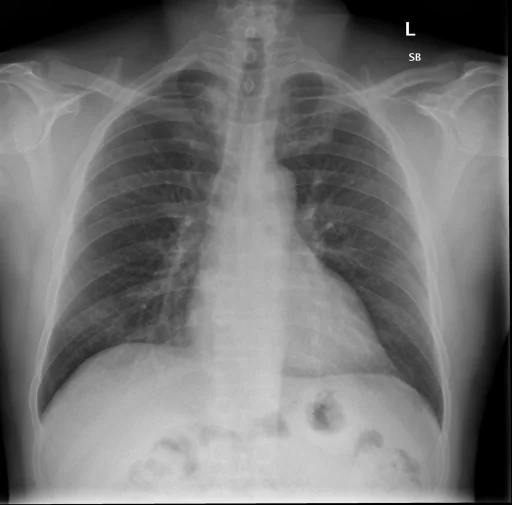
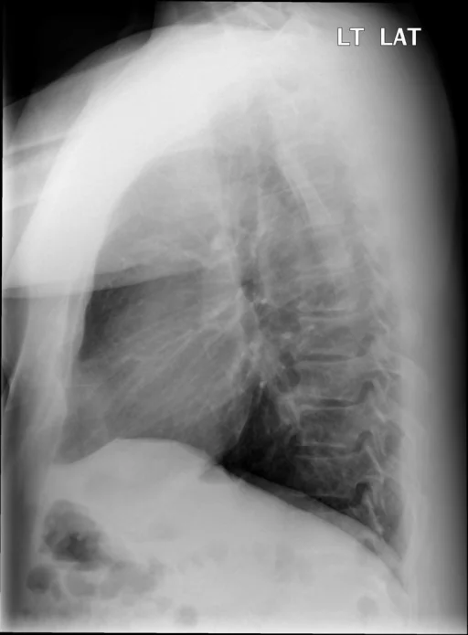
What does this chest radiograph show?
This chest radiograph is classified as “Normal” in the Indiana University Chest X-ray Collection, and no active disease finding is recorded against the case in its structured metadata. That classification rests on the annotation labels recorded in the source dataset, and no active-disease label has been assigned to this case. The structured record behind this page carries a single indexed term; in the source report text, a findings section, an impression and a comparison study are all present. The study comprises 2 views.
This summary is composed from the collection's structured annotation fields, and is not a description of the image.
- Imaging coverage: 2 views, native resolution 512x505 pixels
- Related keywords: Normal, normal, Normal chest radiograph, Chest radiograph and Indiana University Chest X-ray Collection
Radiologist's Diagnostic Impression (analytical summary)
The case is classified as “Normal” and no active disease finding is recorded in its structured metadata.
Case Radiographs (2 Views)
Radiographic Study Specifications
- Case Number
- CXR 502
- Modality
- Chest Radiography (X-Ray)
- Views
- 2 images
- Clinical Status
- Normal
Primary Clinical Report (verbatim source text - NoDerivatives)
- Indication
- The patient is an Adult male with abdominal pain, question of small bowel obstruction. Onset and wheezing in bilateral lung fields.
- Comparison
- Chest, abdominal CT.
- Findings
- Chest. The trachea is midline. Negative for pneumothorax, pleural effusion or focal airspace consolidation. The heart size is normal. Abdomen. No pneumoperitoneum. There is a normal bowel gas pattern. Air and stool visible throughout the entire large colon including the rectum. No abnormally dilated small bowel loops. No evidence for intussusception or small bowel obstruction. No pathologic calcifications over the abdomen or pelvis. Are without fracture or destructive lesion, though there are mild degenerative changes throughout the lumbar spine. Small hiatal hernia is not as well demonstrated on this exam.
- Impression
- Chest. 1. No acute cardiopulmonary abnormality. Abdomen. 1. No acute intra-abdominal process. Negative for obstruction.
Related Radiology Cases
Source, Scientific Provenance and Licence
Indiana University Chest X-ray Collection · Open-i / U.S. National Library of Medicine · CC BY-NC-ND 4.0
Licence: CC BY-NC-ND 4.0
Primary Data Source: https://openi.nlm.nih.gov/