Emphysema / Hyperlucent Lung, Aorta / Thoracic Aorta and Diaphragm / Eventration
What does this chest radiograph show?
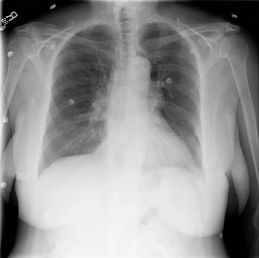
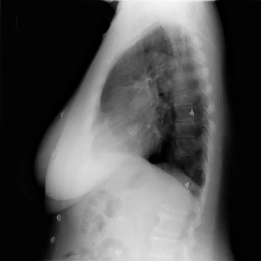
This chest radiograph is indexed in the Indiana University Chest X-ray Collection under “Aorta / Thoracic Aorta, Diaphragm / Eventration and Emphysema / Hyperlucent Lung”. The principal recorded finding is “Emphysema / Hyperlucent Lung”, and the case status is documented as “Active or unspecified chronicity”. Recorded descriptors for this case include laterality Left, and severity Flattened, Mild and Prominent. The structured record behind this page carries 6 indexed terms; in the source report text, a findings section and an impression are present, with no comparison study recorded. The study comprises 2 views.
This summary is composed from the collection's structured annotation fields, and is not a description of the image.
- Involvement site: Left
- Severity and descriptors: Flattened, Mild, Prominent and Tortuous
- Diagnostic category: Emphysema / Hyperlucent Lung, Aorta / Thoracic Aorta, Diaphragm / Eventration, Markings (bronchovascular), Catheters / Tubes / Implanted Devices and Other
- Imaging coverage: 2 views, native resolution 512x510 pixels
- Related keywords: Aorta / Thoracic Aorta, Diaphragm / Eventration, Emphysema / Hyperlucent Lung, Aorta, Diaphragm and Epicardial Fat
Radiologist's Diagnostic Impression (analytical summary)
Recorded findings: Emphysema / Hyperlucent Lung, Aorta / Thoracic Aorta, Diaphragm / Eventration, Aorta (Tortuous, Mild) and Diaphragm (Flattened). Categories: “Aorta / Thoracic Aorta, Diaphragm / Eventration and Emphysema / Hyperlucent Lung”. Status: “Active or unspecified chronicity”.
Case Radiographs (2 Views)
Radiographic Study Specifications
- Case Number
- CXR 867
- Modality
- Chest Radiography (X-Ray)
- Views
- 2 images
- Clinical Status
- Active or unspecified chronicity
Primary Clinical Report (verbatim source text - NoDerivatives)
- Indication
- Adult female, pain
- Findings
- Heart size near top normal, bilateral hilar fullness nonspecific in appearance, mild aortic ectasia/tortuosity. Diaphragm flattening and relative apical lucencies suggestive of emphysema, and irregular interstitial markings, right greater than left. Prominent left epicardial fat, no focal alveolar consolidation, no definite pleural effusion seen. Atrial septal occluder artifact. Mild spine curvature.
- Impression
- Borderline heart size and abnormal interstitial pulmonary pattern which may be compatible with chronic interstitial change, differential diagnosis is and includes asymmetric pulmonary edema, inflammation, atypical infection, infiltrative process. Comparison with previous exams would be of. Bilateral hilar fullness may indicate pulmonary hypertension and clinical correlation is recommended, differential diagnosis reactive lymphadenopathy, metastatic disease.
Related Radiology Cases
Source, Scientific Provenance and Licence
Indiana University Chest X-ray Collection · Open-i / U.S. National Library of Medicine · CC BY-NC-ND 4.0
Licence: CC BY-NC-ND 4.0
Primary Data Source: https://openi.nlm.nih.gov/