Seminoma - Dx path proven
Patient Presentation & Demographics
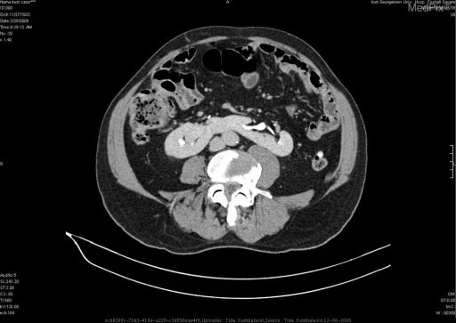
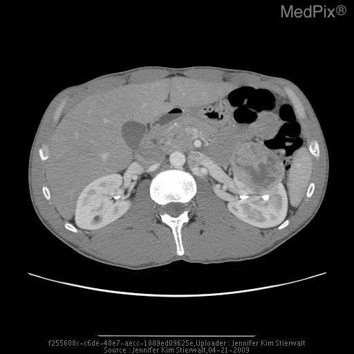
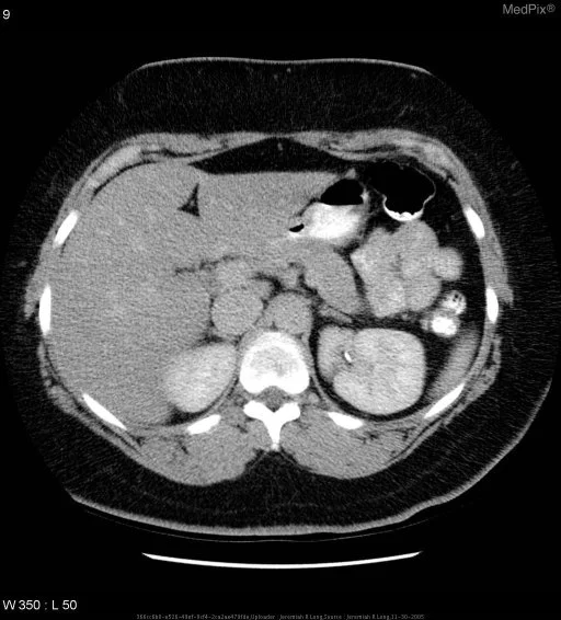
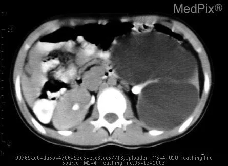
Clinical History & Presentation: Patient presented with characteristic clinical findings consistent with suspected Seminoma - Dx path proven. Multimodal diagnostic imaging was performed to characterize anatomic involvement and guide therapeutic management.
Systematic Imaging Findings
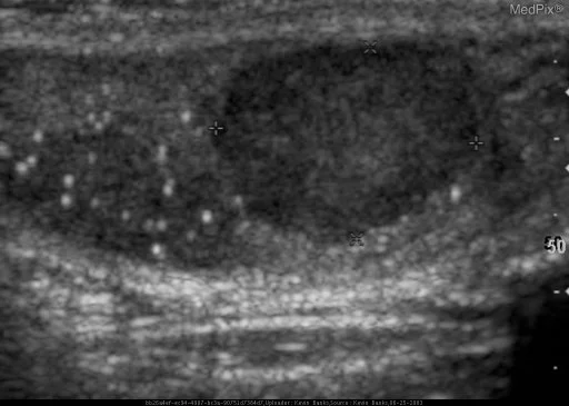
Modality & Anatomic Assessment (Ultrasound (US)): Genitourinary imaging systematically interrogates renal position, bilateral parenchymal thickness, Hounsfield unit density characteristics, pelvicalyceal and ureteral drainage, adrenal gland morphology, and urinary bladder distension with mural uniformity. Key Observational Findings: Sagittal image of the left testis demonstrates two well-defined, hypoechoic lesions as well as diffuse microlithiases. Application of color doppler demonstrates the presence of hypervascular flow within the identified testicular lesions.
Challenge: What Is the Primary Diagnosis?
Evaluate the patient presentation, systematic radiologic findings, and multi-frame imaging series above. Test your clinical reasoning before revealing the definitive diagnosis, hallmark differential criteria, and board pearls.
Seminoma - Dx path proven
Differential Diagnosis (DDx) • Key Clinical Hallmarks
Renal Cell Carcinoma
Distinguishing Radiologic Hallmark: Heterogeneous cortical mass with avid early arterial phase contrast enhancement, rapid washout, and potential renal vein extension.
Angiomyolipoma
Distinguishing Radiologic Hallmark: Cortical renal mass demonstrating macroscopic intracellular fat with negative Hounsfield units (HU < -20) on unenhanced CT.
Complex Bosniak Renal Cyst
Distinguishing Radiologic Hallmark: Cystic renal lesion with thick or irregular septa, nodular mural enhancement, or solid soft tissue components (Bosniak III/IV).
Renal Infarction
Distinguishing Radiologic Hallmark: Wedge-shaped, sharply demarcated non-enhancing renal parenchymal defect with preserved subcapsular rim sign.
High-Yield Clinical Pearls & Management Guidelines
Unenhanced CT attenuation < 10 HU reliably confirms lipid-rich benign adrenal adenoma, eliminating the need for further invasive workup. For complex renal cysts, the revised Bosniak Classification 2019 categorizes lesions based on wall thickness, septal enhancement, and nodularity to direct surgical versus conservative surveillance protocols. ACR Communication Urgency: Tier 2.