Poorly Differentiated Non-Small Cell Lung Carcinoma
Patient Presentation & Demographics
Clinical History & Presentation: Patient presented with characteristic clinical findings consistent with suspected Poorly Differentiated Non-Small Cell Lung Carcinoma. Multimodal diagnostic imaging was performed to characterize anatomic involvement and guide therapeutic management.
Systematic Imaging Findings
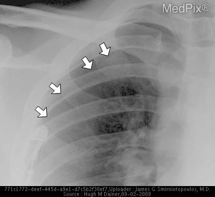
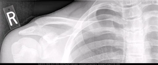
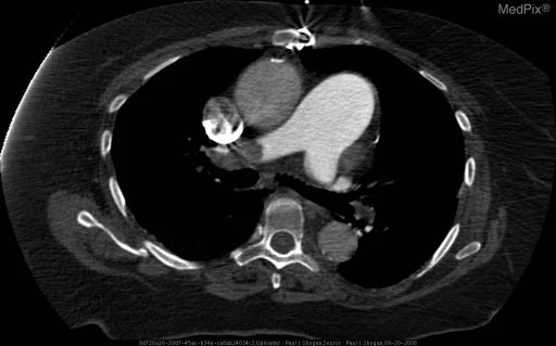
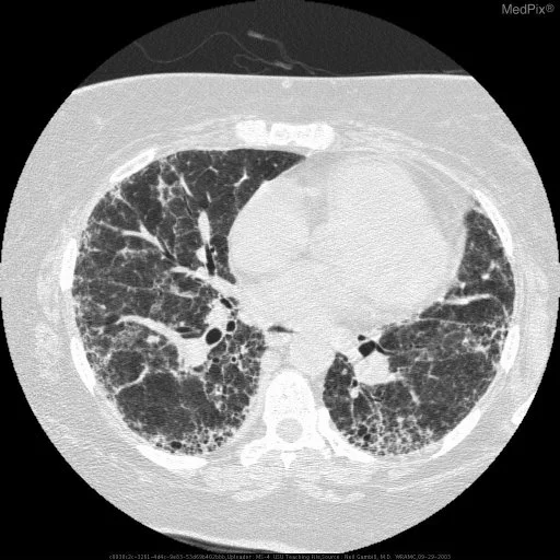
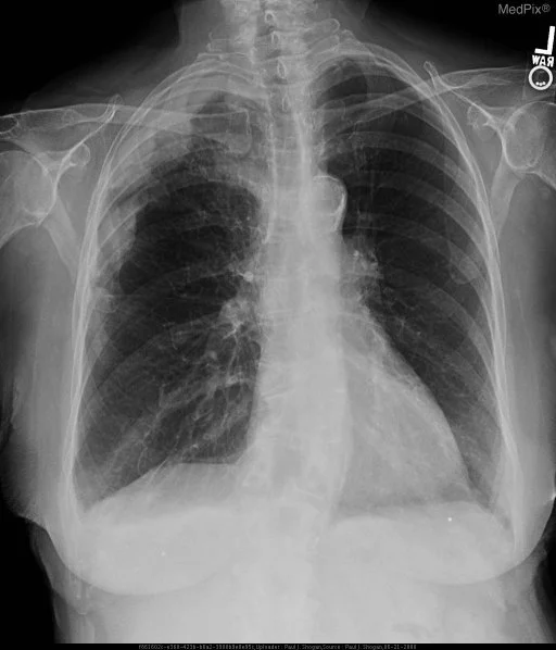
Modality & Anatomic Assessment (Radiography (X-Ray)): Thoracic radiography and CT series systematically evaluate lung parenchymal attenuation, lobar and segmental architecture, tracheobronchial tree patency, pleural surfaces, and cardiomediastinal contours. High-resolution reconstructions identify consolidation, ground-glass opacities, interstitial reticulation, and lymphadenopathy. Key Observational Findings: Dual-energy PA and subtraction soft-tissue-selective radiographs demonstrates nodular, irregular pleural thickening involving the right upper and mid peripheral lung fields. Blunting of the right costophrenic angle is noted. Contrast enhanced axial CT images through various levels of the chest demonstrate a unilateral right sided pleural effusion, with mildly enhancing circumferential, nodular, irregular thickened pleura, encasing the right hemithorax with a rindlike appearance. Additionally, there is focal nodularity of the mediastinal pleura. There is no significant adenopathy, nor evidence of a chest wall abnormality.
Challenge: What Is the Primary Diagnosis?
Evaluate the patient presentation, systematic radiologic findings, and multi-frame imaging series above. Test your clinical reasoning before revealing the definitive diagnosis, hallmark differential criteria, and board pearls.
Poorly Differentiated Non-Small Cell Lung Carcinoma
Differential Diagnosis (DDx) • Key Clinical Hallmarks
Primary Lung Adenocarcinoma
Distinguishing Radiologic Hallmark: Subsolid or spiculated solid pulmonary nodule with pleural indentation, bubble-like lucencies, and vascular convergence.
Bacterial Pneumonia
Distinguishing Radiologic Hallmark: Lobar or segmental air-space consolidation with patent air bronchograms and preservation of underlying parenchymal architecture.
Healed Granuloma
Distinguishing Radiologic Hallmark: Dense central, concentric, or diffuse calcification in a well-defined nodule with documented geometric stability over prior imaging.
Round Atelectasis
Distinguishing Radiologic Hallmark: Curvilinear bronchovascular bundles converging into peripheral pleural-based rounded opacity (comet-tail sign) with adjacent pleural thickening.
High-Yield Clinical Pearls & Management Guidelines
Solitary pulmonary nodule management requires strict adherence to the Fleischner Society Guidelines (2017), utilizing average axial dimensions. The Golden S-curve sign indicates right upper lobe collapse secondary to a central obstructing neoplasm. ACR Communication Urgency: Tier 2 (Action within 6-12 hours; Tier 1 for tension pneumothorax or saddle PE).