Microcystic meningioma
Patient Presentation & Demographics
Clinical History & Presentation: Patient presented with characteristic clinical findings consistent with suspected Microcystic meningioma. Multimodal diagnostic imaging was performed to characterize anatomic involvement and guide therapeutic management.
Systematic Imaging Findings
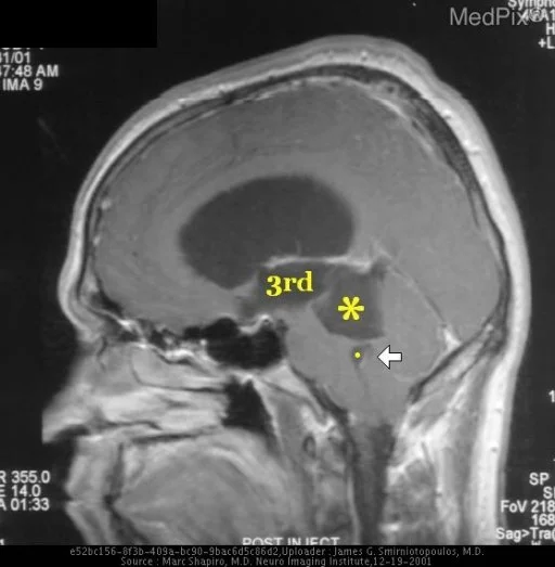
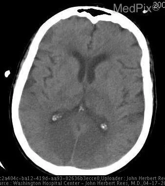
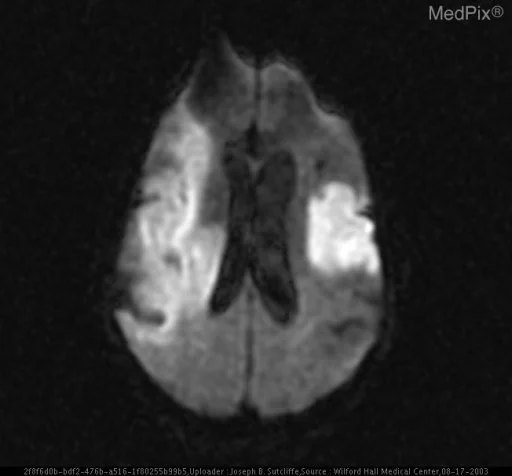
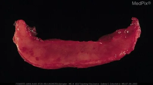
Modality & Anatomic Assessment (Pathology / Histopathology): Cross-sectional neuroimaging series systematically evaluate cerebral and cerebellar parenchyma, grey-white matter differentiation, ventricular size and symmetry, basal cisterns, and extra-axial spaces. Advanced sequences (DWI/ADC, FLAIR, T2*) assess for cytotoxic edema, mass effect, herniation risk, and hemorrhage. Key Observational Findings: Cross section of resected tumor. Note the moist, "humid", cut surface. Microscopic section showing myriads of intercellular and intracellular spaces filled with proteinacous fluid. Note the virtual absence of whorls and psammoma bodies.
Challenge: What Is the Primary Diagnosis?
Evaluate the patient presentation, systematic radiologic findings, and multi-frame imaging series above. Test your clinical reasoning before revealing the definitive diagnosis, hallmark differential criteria, and board pearls.
Microcystic meningioma
Differential Diagnosis (DDx) • Key Clinical Hallmarks
High-Grade Glioma
Distinguishing Radiologic Hallmark: Infiltrative heterogeneously enhancing mass with central necrosis, thick irregular rim enhancement, and extensive surrounding vasogenic edema.
Brain Metastasis
Distinguishing Radiologic Hallmark: Well-circumscribed lesion at grey-white matter junction with disproportionately extensive vasogenic edema relative to lesion size.
Pyogenic Brain Abscess
Distinguishing Radiologic Hallmark: Thin, smooth, uniform enhancing rim showing intense central diffusion restriction (bright on DWI, dark on ADC) and hypointense T2 capsule.
Tumefactive Demyelinating Lesion
Distinguishing Radiologic Hallmark: Large demyelinating plaque with incomplete rim enhancement (open-ring sign) opening toward the cerebral cortex without significant mass effect.
High-Yield Clinical Pearls & Management Guidelines
In ring-enhancing intracranial lesions, intense central diffusion restriction on DWI with matching low ADC values reliably confirms pyogenic brain abscess versus necrotic neoplasm. Non-contrast head CT remains the initial gold standard for hyperacute intracranial hemorrhage. ACR Communication Urgency: Tier 1 (Immediate notification within 30-60 minutes for herniation or acute hemorrhage).