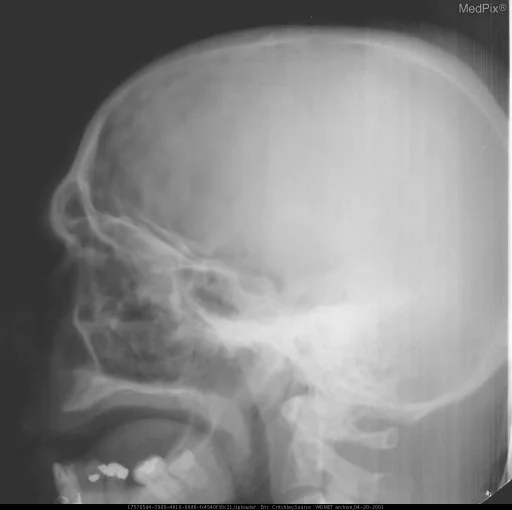
WDMET radiographs
Patient Presentation & Demographics
Clinical History & Presentation: Patient presented with characteristic clinical findings consistent with suspected WDMET radiographs. Multimodal diagnostic imaging was performed to characterize anatomic involvement and guide therapeutic management.
Systematic Imaging Findings
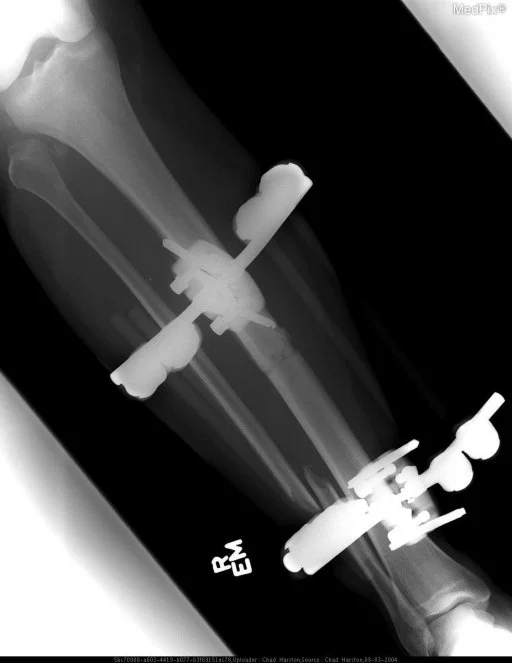
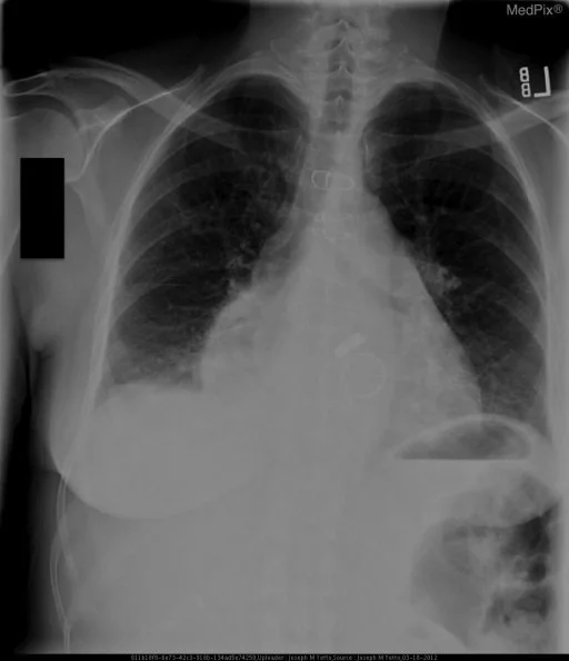
Modality & Anatomic Assessment (Radiography (X-Ray)): Musculoskeletal series systematically evaluate cortical bone integrity, trabecular architecture, joint space width, subchondral bone mineral density, and adjacent soft-tissue envelopes. Multiplanar projections detect occult fractures, periosteal reactions, and articular alignment discrepancies.
Challenge: What Is the Primary Diagnosis?
Evaluate the patient presentation, systematic radiologic findings, and multi-frame imaging series above. Test your clinical reasoning before revealing the definitive diagnosis, hallmark differential criteria, and board pearls.
WDMET radiographs
Differential Diagnosis (DDx) • Key Clinical Hallmarks
Erosive Osteoarthritis
Distinguishing Radiologic Hallmark: Non-uniform joint space narrowing with subchondral sclerosis, osteophyte formation, and gull-wing gullies in interphalangeal joints.
Rheumatoid Arthritis
Distinguishing Radiologic Hallmark: Symmetric marginal erosions, periarticular osteopenia, and uniform joint space loss without osteophyte proliferation.
Acute Osteomyelitis
Distinguishing Radiologic Hallmark: Permeative cortical bone destruction, multilayered periosteal reaction, subperiosteal abscess, and devitalized sequestrum formation.
Osteosarcoma
Distinguishing Radiologic Hallmark: Aggressive periosteal reaction (Codman triangle, sunburst appearance) with cloud-like osteoid tumor matrix and soft tissue extension.
High-Yield Clinical Pearls & Management Guidelines
In elderly patients presenting with hip pain after low-energy trauma and negative initial radiographs, immediate non-contrast MRI or CT is indicated to exclude occult femoral neck fracture. The "fallen fragment sign" in a lytic bone lesion is pathognomonic for a pathologic unicameral bone cyst. ACR Communication Urgency: Tier 2 for unstable trauma, Tier 3 for routine staging.