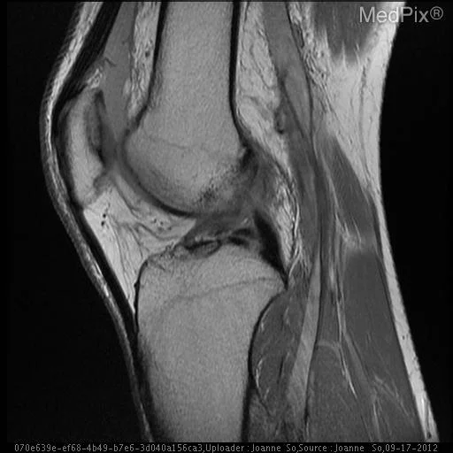
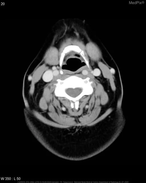
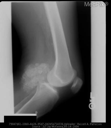
Giant Cell Bone Tumor
Patient Presentation & Demographics
Clinical History & Presentation: Patient presented with characteristic clinical findings consistent with suspected Giant Cell Bone Tumor. Multimodal diagnostic imaging was performed to characterize anatomic involvement and guide therapeutic management.
Systematic Imaging Findings
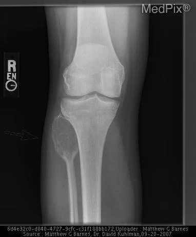
Modality & Anatomic Assessment (Radiography (X-Ray)): Musculoskeletal series systematically evaluate cortical bone integrity, trabecular architecture, joint space width, subchondral bone mineral density, and adjacent soft-tissue envelopes. Multiplanar projections detect occult fractures, periosteal reactions, and articular alignment discrepancies. Key Observational Findings: Anteroposterior and lateral radiographs of the right knee demonstrate an expansile lytic lesion of the head of the proximal fibula with fairly narrow zone of transition. No associated periosteal reaction or soft tissue mass. Tibia and femur are normal in appearance. Joint spaces are maintained. No joint effusion. Tunnel radiograph of the right knee demonstrates an expansile lytic lesion of the proximal fibula with fairly narrow zone of transition. No associated periosteal reaction or soft tissue mass. Tibia and femur are normal in appearance. Joint spaces are maintained. No joint effusion.
Challenge: What Is the Primary Diagnosis?
Evaluate the patient presentation, systematic radiologic findings, and multi-frame imaging series above. Test your clinical reasoning before revealing the definitive diagnosis, hallmark differential criteria, and board pearls.
Giant Cell Bone Tumor
Differential Diagnosis (DDx) • Key Clinical Hallmarks
Erosive Osteoarthritis
Distinguishing Radiologic Hallmark: Non-uniform joint space narrowing with subchondral sclerosis, osteophyte formation, and gull-wing gullies in interphalangeal joints.
Rheumatoid Arthritis
Distinguishing Radiologic Hallmark: Symmetric marginal erosions, periarticular osteopenia, and uniform joint space loss without osteophyte proliferation.
Acute Osteomyelitis
Distinguishing Radiologic Hallmark: Permeative cortical bone destruction, multilayered periosteal reaction, subperiosteal abscess, and devitalized sequestrum formation.
Osteosarcoma
Distinguishing Radiologic Hallmark: Aggressive periosteal reaction (Codman triangle, sunburst appearance) with cloud-like osteoid tumor matrix and soft tissue extension.
High-Yield Clinical Pearls & Management Guidelines
In elderly patients presenting with hip pain after low-energy trauma and negative initial radiographs, immediate non-contrast MRI or CT is indicated to exclude occult femoral neck fracture. The "fallen fragment sign" in a lytic bone lesion is pathognomonic for a pathologic unicameral bone cyst. ACR Communication Urgency: Tier 2 for unstable trauma, Tier 3 for routine staging.