Intravascular lymphomatosis (IVL)
Patient Presentation & Demographics
Clinical History & Presentation: Patient presented with characteristic clinical findings consistent with suspected Intravascular lymphomatosis (IVL). Multimodal diagnostic imaging was performed to characterize anatomic involvement and guide therapeutic management.
Systematic Imaging Findings
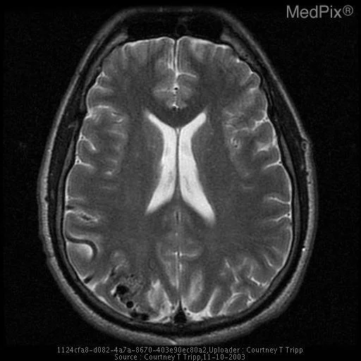
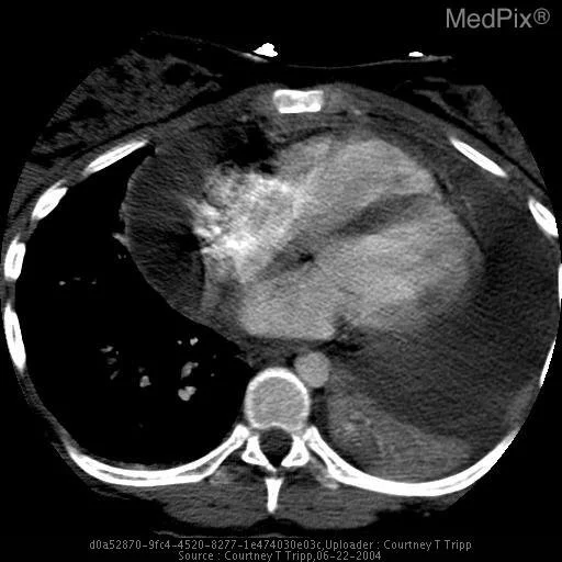
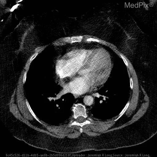
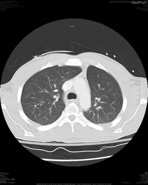
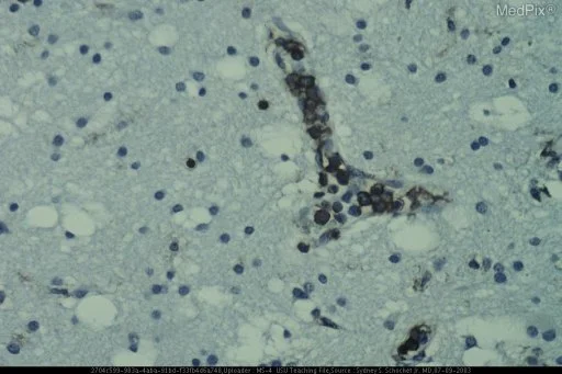
Modality & Anatomic Assessment (Pathology / Histopathology): Cross-sectional cardiovascular imaging systematically evaluates thoracic aortic caliber (ascending, arch, descending), mediastinal contour, pericardial space, myocardial thickness, ventricular chambers, and major pulmonary arterial branches. Contrast-enhanced phases differentiate luminal filling defects, intramural collections, and dissection flap extent. Key Observational Findings: A section of the biopsy specimen stained for leukocyte common antigen shows staining of the intralumenal neoplastic cells. Coronal section of the brain showing small focally necrotic lesions in the white matter of both hemispheres.
Challenge: What Is the Primary Diagnosis?
Evaluate the patient presentation, systematic radiologic findings, and multi-frame imaging series above. Test your clinical reasoning before revealing the definitive diagnosis, hallmark differential criteria, and board pearls.
Intravascular lymphomatosis (IVL)
Differential Diagnosis (DDx) • Key Clinical Hallmarks
Stanford Type A Aortic Dissection
Distinguishing Radiologic Hallmark: Intimal flap visualization within ascending aorta lumen separating true and false lumens, with potential extension into aortic arch vessels.
Thoracic Aortic Aneurysm
Distinguishing Radiologic Hallmark: Fusiform or saccular aortic caliber dilatation exceeding 50% of reference normal diameter without internal intimal flap.
Aortic Intramural Hematoma
Distinguishing Radiologic Hallmark: Crescentic non-enhancing hyperattenuating thickening within the aortic wall without detectable intimal flap or flow communication.
Acute Pulmonary Embolism
Distinguishing Radiologic Hallmark: Intraluminal filling defect within pulmonary arterial branches with secondary RV/LV diameter ratio exceeding 1.0 indicating right heart strain.
High-Yield Clinical Pearls & Management Guidelines
Stanford Type A aortic dissection involves the ascending aorta and represents a critical surgical emergency requiring immediate open cardiovascular repair. In acute pulmonary embolism, an RV/LV diameter ratio > 1.0 on transverse CT reconstructions indicates acute right ventricular strain and high short-term adverse outcome risk. ACR Communication Urgency: Tier 1 (Action within 30-60 minutes).