Comprehensive 7-Step Electrophysiological Interpretation
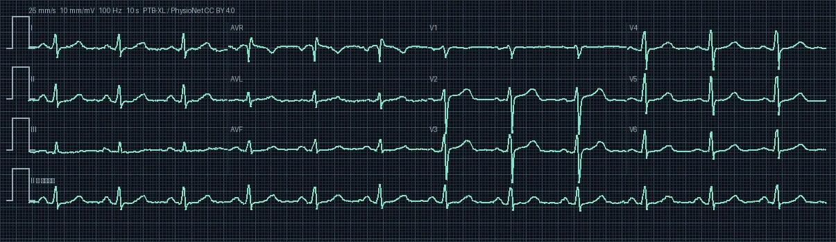
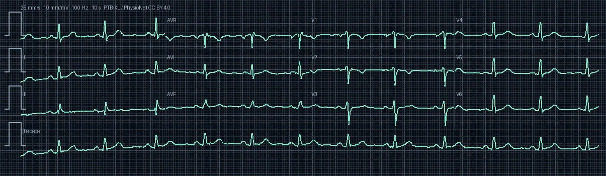
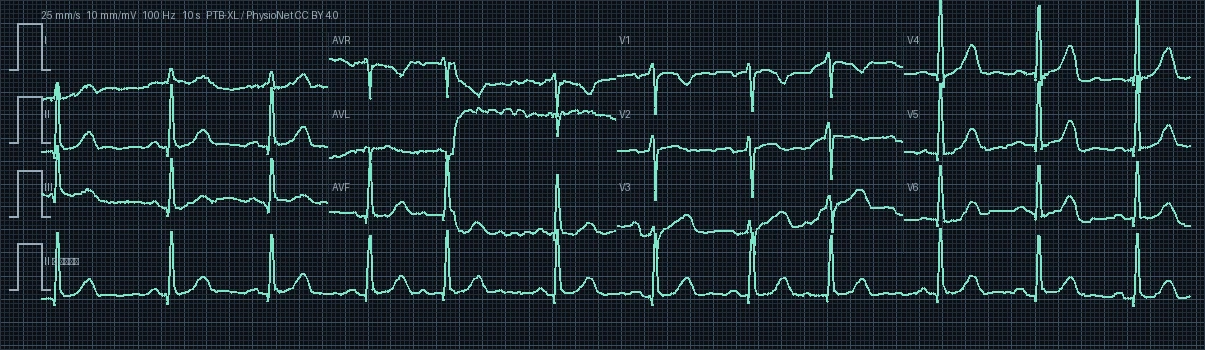
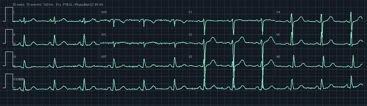
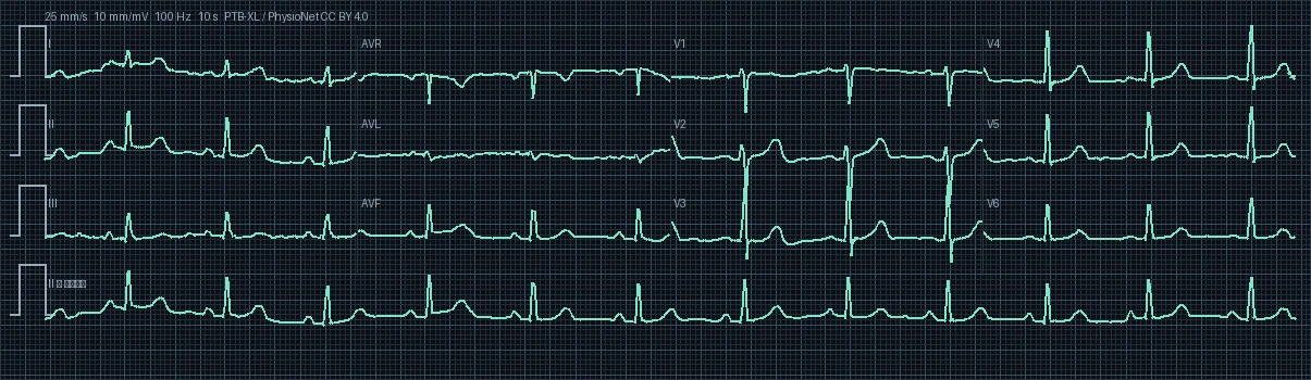
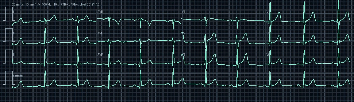
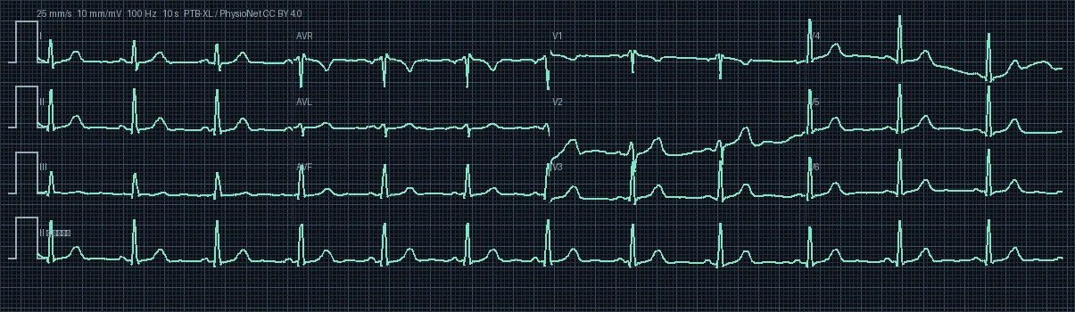
The electrocardiogram displays Normal Sinus Rhythm (NSR) with steady regular sequence and intact 1:1 atrioventricular relationship. Cardiac activation initiates normally within the sinoatrial (SA) node in the high posterior right atrium, spreading through the internodal pathways to the AV node. P waves are distinct, uniform, and upright in inferior leads.
The resting ventricular heart rate is quantified at 73.2 beats per minute (bpm) with a median R-R interval of 820 ms. This rate is within the physiologic normocardia range (60 to 100 bpm), reflecting balanced resting sympathovagal tone and stable cardiac output.
The mean frontal plane QRS electrical axis is calculated at 121.5° (Right Axis Deviation (RAD)). Displays Right Axis Deviation (+90° to +180°). The rightward shift points toward right ventricular pressure/volume overload (RVH), chronic obstructive pulmonary disease, pulmonary embolism, or left posterior fascicular block.
Electrophysiologic interval analysis: PR interval is normal at 160 ms (normal range 120-200 ms), confirming physiological transmission velocity across the atrioventricular node. QRS complex is narrow at 110 ms (<100 ms), validating rapid, synchronized biventricular activation via the specialized His-Purkinje network. Bazett-corrected QTc (QTc = QT / √RR) is measured at 452.8 ms (raw QT: 410 ms), resting comfortably within safe gender-specific limits for females and confirming stable ventricular repolarization.
P-wave morphology across all 12 leads is unremarkable, without evidence of right or left atrial enlargement. Precordial R-wave progression from lead V1 to V6 is physiological with a normal transition zone at V3-V4. ST segments remain strictly isoelectric throughout, devoid of pathologic elevations or depressions, and T waves exhibit normal upright symmetry concordant with QRS vectors.
Biophysical clinical correlation: In this 49-year-old female patient, electrocardiographic parameters must be framed within adult cardiovascular risk profiles (hypertension, metabolic profile, and lifestyle); intact intervals and isoelectric baselines reflect resting myocardial stability. Furthermore, calculated QTc intervals and resting heart rate closely align with metabolic perfusion demands for this demographic group.
Key differential diagnoses for this normal tracing include: 1. Normal physiologic variant 2. Benign early repolarization variant 3. Transient autonomic fluctuations secondary to anxiety or hyperventilation 4. Subclinical coronary artery disease (normal at rest, requiring stress testing if symptomatic) 5. Minor electrode positioning artifact.