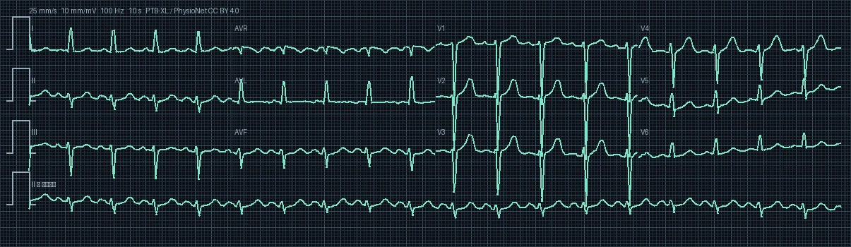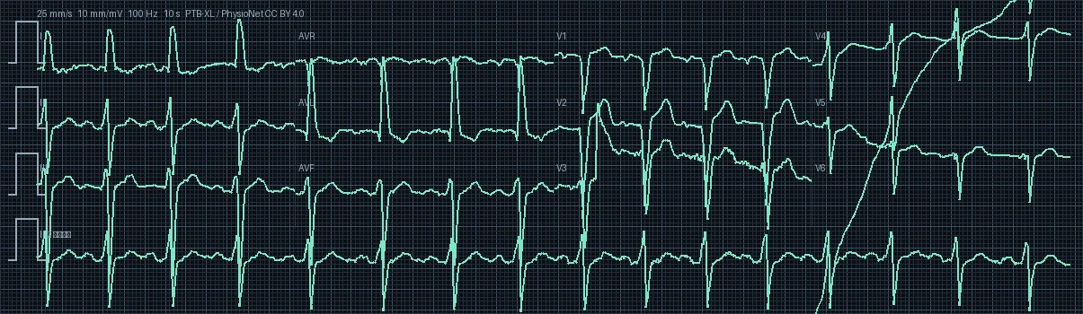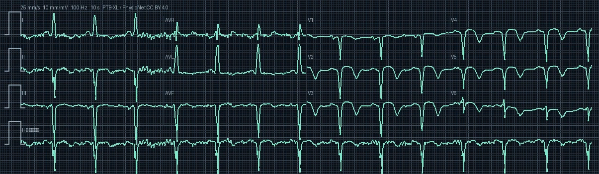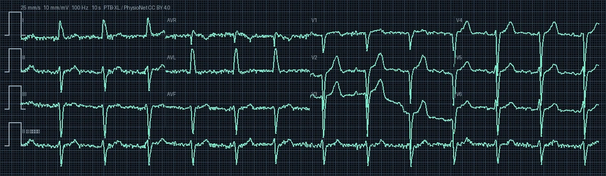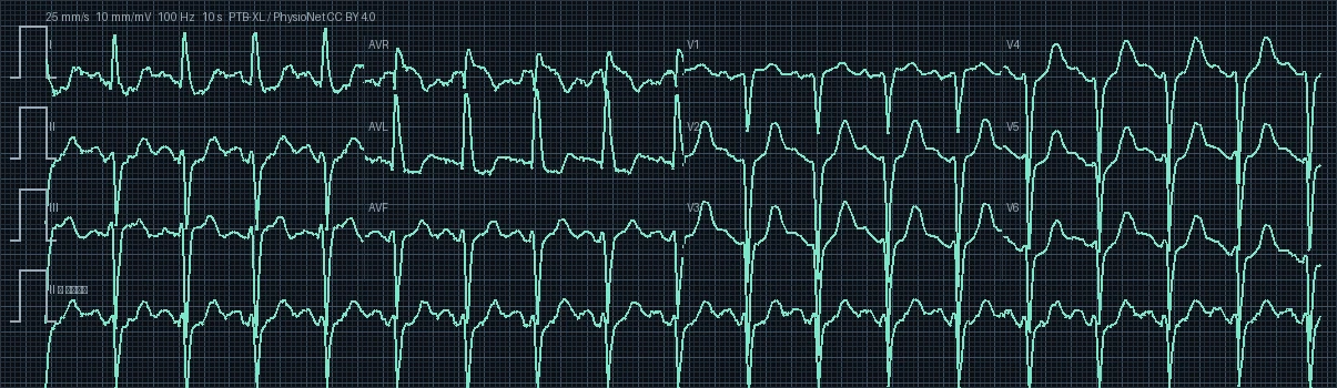
ECG #924 — Anterolateral myocardial infarction

PTB-XL Record #00924 · 12-Lead Diagnostic Electrocardiogram · 500 Hz High-Resolution

12-Lead Electrocardiogram Preview #924
Target Territory: Anterolateral Territory
I II III aVR aVL aVF V1 V2 V3 V4 V5 V6
Heart Rate
109.1 bpm
PR Interval
—
QRS Duration
110 ms
QTc (Bazett)
445.0 ms
Frontal QRS Axis
-76.8° (Left Axis Deviation)
Demographics
88 yrs · Female

Clinical Direct Answer

The primary diagnostic finding on this 12-lead electrocardiogram is anterolateral myocardial infarction, recorded with an underlying rhythm of sinus tachycardia at a rate of 109.1 beats per minute (tachycardia). Pathological waveform alterations are predominantly localized to target territory leads, requiring prompt clinical correlation, serial biomarkers, and targeted electrophysiological evaluation.

Comprehensive 7-Step Electrophysiological Interpretation

1

Rhythm & Pacemaker Origin

The tracing exhibits Sinus Tachycardia with preserved regular rhythmicity and 1:1 AV association. Impulse formation originates in the sinoatrial node driven by enhanced sympathetic adrenergic tone or physiologic stress. Uniform upright P waves consistently precede each QRS complex.
2

Ventricular Rate & Regularity

The resting ventricular heart rate is quantified at 109.1 beats per minute (bpm) with a median R-R interval of 550 ms. This rate is exceeding 100 bpm, categorizing this record as sinus tachycardia, which attenuates coronary perfusion time during shortened diastole.

3

Frontal Plane QRS Axis & Vector Analysis

The mean frontal plane QRS electrical axis is calculated at -76.8° (Left Axis Deviation (LAD)). Displays Left Axis Deviation (-30° to -90°). Depolarization is directed superiorly and leftward, consistent with left anterior fascicular block (LAFB), left ventricular hypertrophy, or inferior wall scar.

4

Conduction Intervals & Chamber Timing (PR, QRS, QT/QTc)

Electrophysiologic interval analysis: PR interval is non-measurable due to fibrillatory baseline activity or absence of discrete P waves. QRS complex is narrow at 110 ms (<100 ms), validating rapid, synchronized biventricular activation via the specialized His-Purkinje network. Bazett-corrected QTc (QTc = QT / √RR) is measured at 445.0 ms (raw QT: 330 ms), resting comfortably within safe gender-specific limits for females and confirming stable ventricular repolarization.

5

Waveform Morphology & ST-T Segment Dynamics

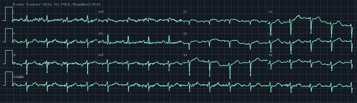
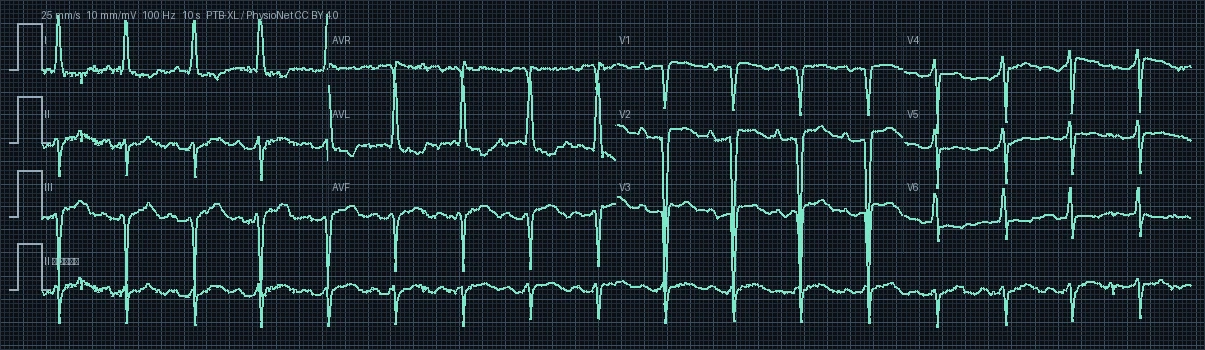
Waveform morphology demonstrates classic diagnostic ischemic patterns corresponding to anterolateral myocardial infarction. Pathological ST-segment deviations accompanied by dynamic T-wave evolution and diagnostic Q waves point toward coronary territorial injury, supported by reciprocal ST depressions in opposite leads.

6

Clinical Correlation & Pathophysiological Significance

Biophysical clinical correlation: Considering this patient's age of 88 years (female), age-associated degenerative fibrosis of the cardiac skeleton and conduction tissue is common; preserved conduction intervals and absence of advanced AV blocks confirm satisfactory resting conduction system integrity. Furthermore, calculated QTc intervals and resting heart rate closely align with metabolic perfusion demands for this demographic group.

7

Differential Diagnosis & Recommended Diagnostic Workup

Key differential diagnoses include: 1. Acute Pericarditis (diffuse concave ST elevations with PR depressions and no reciprocal changes) 2. Benign Early Repolarization (BER) 3. Brugada syndrome in precordial leads V1-V2 4. Takotsubo stress cardiomyopathy 5. Severe Hyperkalemia.

Interactive 12-Lead Signal & Digital Caliper

Select individual leads to examine high-resolution waveforms and measure precise intervals in milliseconds.

Click two points on the tracing to measure interval
Clinical & Educational Notice: This electrocardiogram analysis is generated from the PTB-XL open clinical cohort for educational, reference, and computational research purposes. It does not replace individualized bedside clinical evaluation, emergency triage, or formal cardiologist consultation.