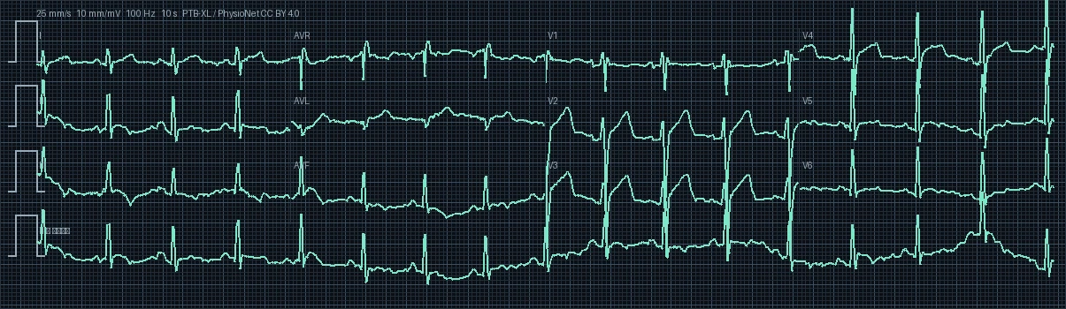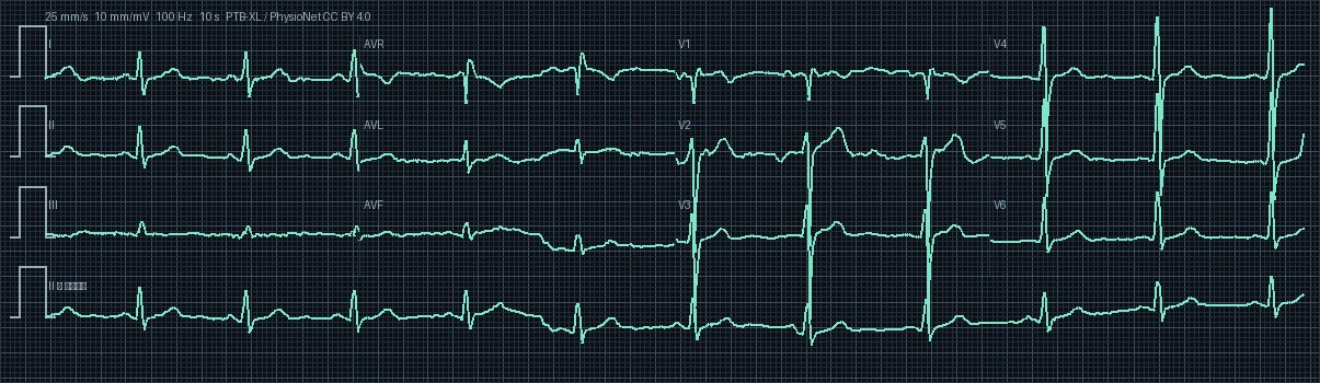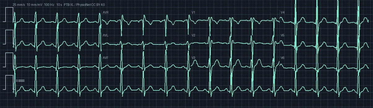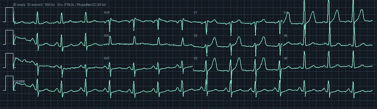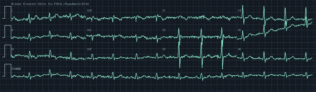
ECG #689 — Incomplete right bundle branch block

PTB-XL Record #00689 · 12-Lead Diagnostic Electrocardiogram · 500 Hz High-Resolution

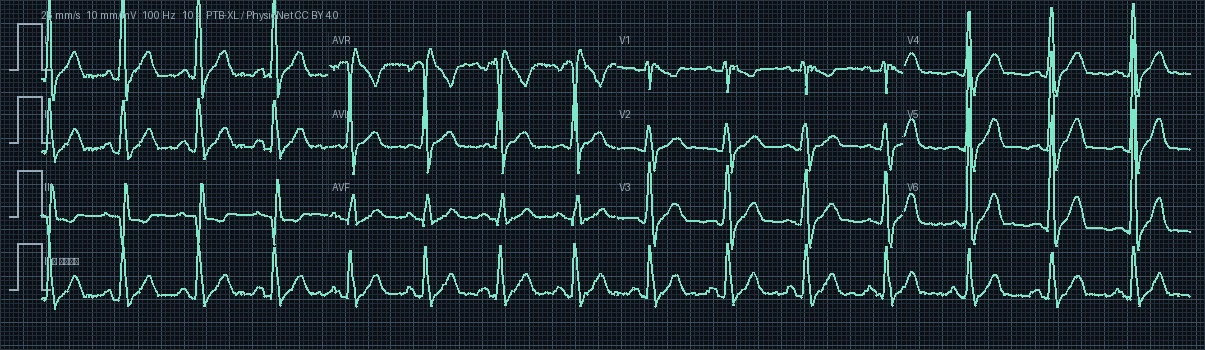
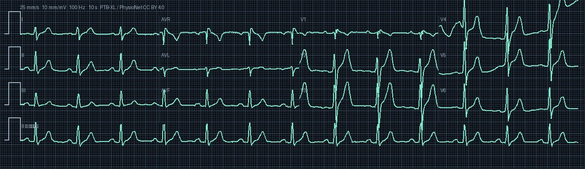
12-Lead Electrocardiogram Preview #689
Target Territory: Incomplete Right Bundle Delay
I II III aVR aVL aVF V1 V2 V3 V4 V5 V6
Heart Rate
87.0 bpm
PR Interval
—
QRS Duration
110 ms
QTc (Bazett)
517.7 ms
Frontal QRS Axis
+144.9° (Right Axis Deviation)
Demographics
Unspecified · Male

Clinical Direct Answer

The primary diagnostic finding on this 12-lead electrocardiogram is incomplete right bundle branch block, recorded with an underlying rhythm of sinus rhythm at a rate of 87.0 beats per minute (normal rate). Pathological waveform alterations are predominantly localized to target territory leads, requiring prompt clinical correlation, serial biomarkers, and targeted electrophysiological evaluation.

Comprehensive 7-Step Electrophysiological Interpretation

1

Rhythm & Pacemaker Origin

The electrocardiogram displays Normal Sinus Rhythm (NSR) with steady regular sequence and intact 1:1 atrioventricular relationship. Cardiac activation initiates normally within the sinoatrial (SA) node in the high posterior right atrium, spreading through the internodal pathways to the AV node. P waves are distinct, uniform, and upright in inferior leads.
2

Ventricular Rate & Regularity

The resting ventricular heart rate is quantified at 87.0 beats per minute (bpm) with a median R-R interval of 690 ms. This rate is within the physiologic normocardia range (60 to 100 bpm), reflecting balanced resting sympathovagal tone and stable cardiac output.

3

Frontal Plane QRS Axis & Vector Analysis

The mean frontal plane QRS electrical axis is calculated at 144.9° (Right Axis Deviation (RAD)). Displays Right Axis Deviation (+90° to +180°). The rightward shift points toward right ventricular pressure/volume overload (RVH), chronic obstructive pulmonary disease, pulmonary embolism, or left posterior fascicular block.

4

Conduction Intervals & Chamber Timing (PR, QRS, QT/QTc)

Electrophysiologic interval analysis: PR interval is non-measurable due to fibrillatory baseline activity or absence of discrete P waves. QRS complex is narrow at 110 ms (<100 ms), validating rapid, synchronized biventricular activation via the specialized His-Purkinje network. Bazett-corrected QTc (QTc = QT / √RR) is prolonged at 517.7 ms above the gender-adjusted normal threshold for males (450 ms), warranting clinical surveillance for polymorphic ventricular tachycardia and electrolyte evaluation.

5

Waveform Morphology & ST-T Segment Dynamics

Precordial and frontal QRS morphology reveals diagnostic bundle branch delay (incomplete right bundle branch block). Notched wide QRS deflections coupled with secondary ST-segment and T-wave repolarization discordance reflect sequential rather than simultaneous biventricular depolarization.

6

Clinical Correlation & Pathophysiological Significance

Biophysical clinical correlation: Patient age is unspecified in the clinical referral record. Electrophysiological interval analysis confirms preserved atrioventricular and intraventricular conduction velocities with normal His-Purkinje integrity. Resting heart rate and rate-corrected repolarization dynamics (QTc) remain physiologically stable without evidence of age-associated conduction defects or degenerative tissue fibrosis.

7

Differential Diagnosis & Recommended Diagnostic Workup

Key differential diagnoses include: 1. Ventricular Paced Rhythm 2. Wolff-Parkinson-White pre-excitation syndrome (short PR with delta wave) 3. Ventricular Tachycardia 4. Non-specific intraventricular conduction delay (IVCD) 5. Sodium-channel blocker toxicity.

Interactive 12-Lead Signal & Digital Caliper

Select individual leads to examine high-resolution waveforms and measure precise intervals in milliseconds.

Click two points on the tracing to measure interval
Clinical & Educational Notice: This electrocardiogram analysis is generated from the PTB-XL open clinical cohort for educational, reference, and computational research purposes. It does not replace individualized bedside clinical evaluation, emergency triage, or formal cardiologist consultation.