Juvenile Xanthogranuloma (JXG)
Diagnostic correlation matching established clinical dermatology and dermatopathology reference guidelines.
What Are the Clinical Hallmarks and Diagnostic Features of Juvenile Xanthogranuloma (JXG)?
Juvenile xanthogranuloma is the most common benign non-Langerhans cell histiocytosis of infancy and childhood with characteristic Touton giant cells.
- Pathologic Category: Inflammatory & Papulosquamous Dermatoses
- Primary Diagnosis: Juvenile Xanthogranuloma (Non-Langerhans Cell Histiocytosis)
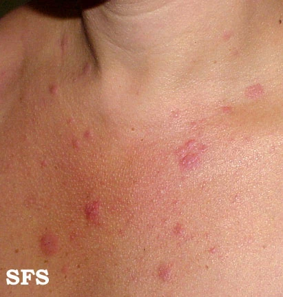
- Clinical Morphology: Firm, solitary dome-shaped yellow-orange to reddish-brown papule or nodule on head and neck.
- High-Resolution Images: 31 standardized dermoscopic and macroscopic photographs
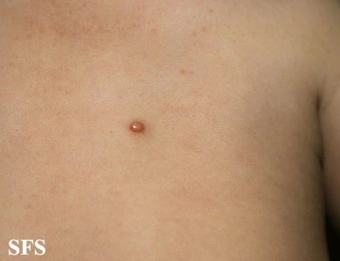
Morphology & Clinical Distribution
Firm, solitary dome-shaped yellow-orange to reddish-brown papule or nodule on head and neck.
Clinical Dermatology Pearls & Biopsy Stewardship
Ophthalmology slit-lamp exam to rule out iris xanthogranuloma; histopathology reveals pathognomonic Touton giant cells.
Clinical Differential Diagnoses (DDx)
Langerhans Cell Histiocytosis
Distinguishing Feature: Ulcerated eczematous lesions, CD1a and Langerin positive.
Solitary Mastocytoma
Distinguishing Feature: Positive Darier sign with urtication upon mechanical stroking.
Molluscum Contagiosum
Distinguishing Feature: Umbilicated dome-shaped translucent papule.
Spitz Nevus
Distinguishing Feature: Erythematous spindle-cell melanocytic lesion.
Case Metadata & Archival Lineage
- Case ID
- 06ae08b7c965add4
- Clinical Category
- Inflammatory & Papulosquamous Dermatoses
- Available Images
- 31 Diagnostic Photographs
- Canonical Slug
- /derm/inflammatory/juvenile-xanthogranuloma/
- Cloud CDN Serving
- cdn.dexamedai.com (100% First-Party)
Clinically Correlated Educational Cases
Academic Attribution & Data Provenance
Dataset: Fitzpatrick17k dataset (Groh et al., 2021) / Atlas Dermatologico
License: Creative Commons Attribution-NonCommercial-ShareAlike 3.0 (CC BY-NC-SA 3.0)